. 2020 Jul 20;9:e56523. doi: 10.7554/eLife.56523

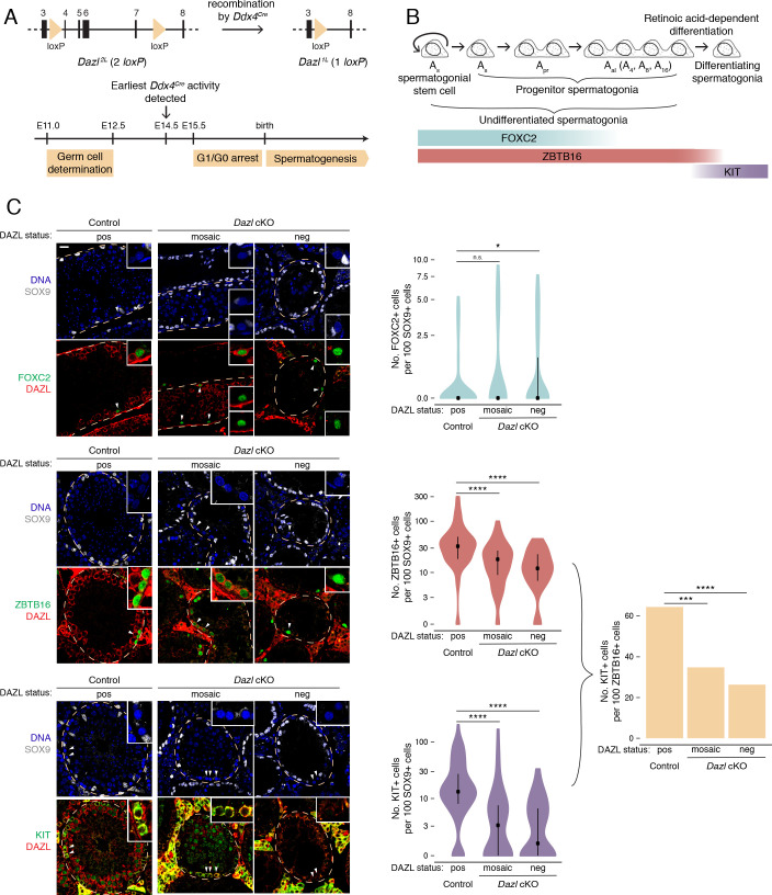
Figure 1. DAZL promotes spermatogonial expansion and differentiation.

(A) Schematic of Dazl2L (two loxP) and Dazl1L (one loxP) alleles. Recombination of Dazl2L allele via Ddx4Cre allele yields Dazl1L allele in germ cells. (B) Schematic of spermatogenesis from Asingle (As) spermatogonial stem cells to progenitor spermatogonia to differentiating spermatogonia, with expression of spermatogonial markers. As spermatogonial stem cells self-renew while also giving rise to As progenitor spermatogonia, which differentiate without self-renewal. Subsequent divisions with incomplete cytokinesis produce chains of two (Apaired, Apr) as well as chains of four, eight, and sixteen (Aaligned, Aal) progenitor spermatogonia. Progenitors form differentiating spermatogonia in response to retinoic acid. In addition, chains of Apr and Aal progenitors can fragment to form As spermatogonial stem cells, particularly under conditions of stress (not shown). (C) Quantification of FOXC2, ZBTB16, and KIT-positive spermatogonia (green) in histological sections of control and Dazl cKO adult testes. SOX9 marks Sertoli cells (gray), DAZL is in red, and DAPI marks DNA (blue). Select tubules are outlined via dotted orange line. Cells enlarged within insets are highlighted by arrowheads. Insets show spermatogonia that are DAZL-positive and FOXC2-positive (control and mosaic cKO panels); DAZL-negative and FOXC2-positive (mosaic and DAZL-negative cKO panels); DAZL-positive and ZBTB16-positive (control and mosaic cKO panels); DAZL-negative and ZBTB16-positive (DAZL-negative cKO panel); DAZL-positive and KIT-positive (control and mosaic cKO panels); and DAZL-negative and KIT-negative (DAZL-negative cKO panel). Scale bar = 20 μm. Populations were quantified from 50 tubules per animal, with two animals per genotype. Violin plots display medians with interquartile ranges. Difference between normalized FOXC2, ZBTB16, and KIT-positive populations was statistically assessed via two-sided Mann-Whitney U test. Difference between normalized KIT-positive populations over normalized ZBTB16-positive populations was statistically assessed via two-sided odds ratio. *, p<0.05; ***, p<0.001; ****, p<0.0001.

Figure 1—source data 1. RNA-seq analysis of Pou5f1:EGFP-positive spermatogonia from control and Dazl cKO testes.
Figure 1—source data 2. Quantification of spermatogonial subpopulations and tubule cross-section area in control and Dazl cKO testes.

Figure 1.

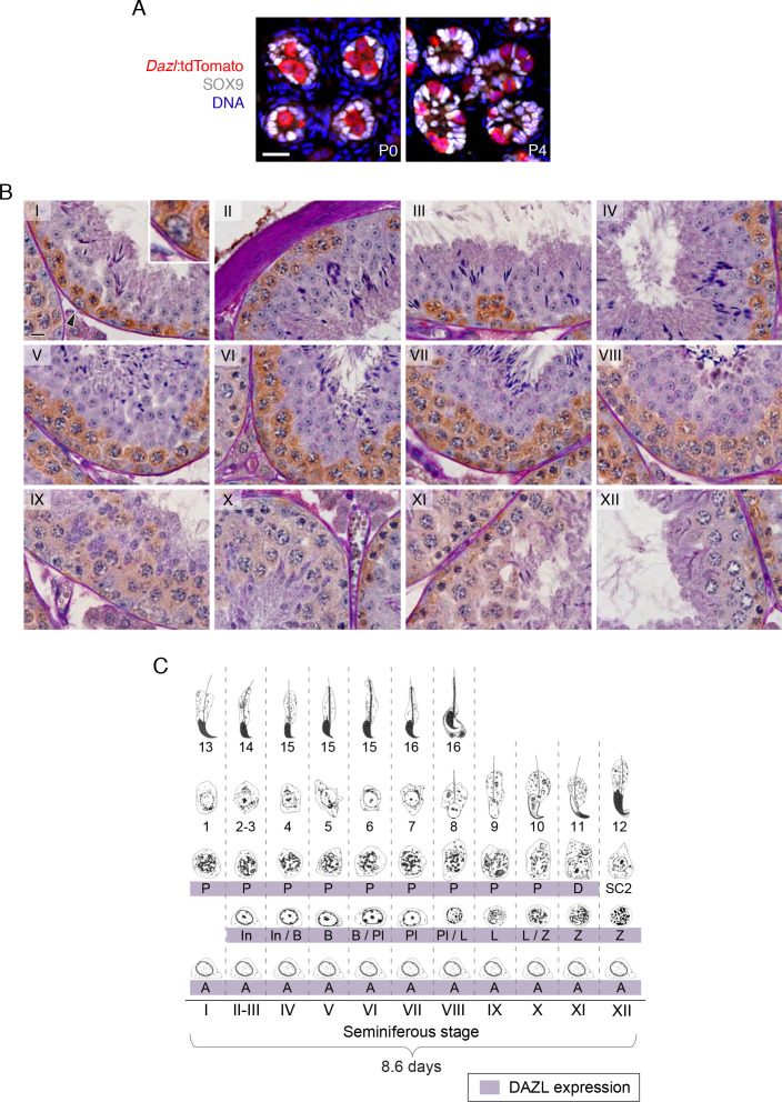
Figure 1—figure supplement 1. DAZL expression in postnatal gonocytes and during spermatogenesis.

Figure 1—figure supplement 1.

(A) Male gonocytes at postnatal days (P) 0 and 4 express the Dazl:tdTomato reporter. SOX9 marks Sertoli cells. Scale bar = 20 μm. (B) DAZL immunohistochemistry in adult testis. DAZL localized to type A, intermediate, and type B spermatogonia as well as spermatocytes from the preleptotene through diplotene stages. Roman numeral designates stage of the primary tubule in each panel. Type A spermatogonium highlighted by arrowhead in stage I tubule is enlarged within inset. Scale bar = 10 μm. (C) Summary of DAZL expression by stage of spermatogenesis, based on immunohistochemistry in B. Abbreviations, in spermatogenic order: A, type A spermatogonium; In, intermediate spermatogonium; B, type B spermatogonium; Pl, preleptotene spermatocyte; L, leptotene spermatocyte; Z, zygotene spermatocyte; D, diplotene spermatocyte; SC2, secondary spermatocytes. The numbers indicate the ‘step’ staging designation of the corresponding spermatids.
Figure 1—figure supplement 2. Conditional deletion of Dazl in spermatogonia.
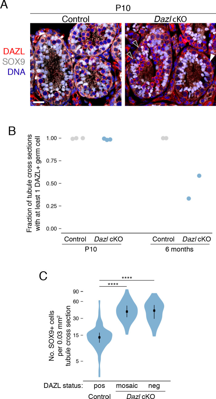
Figure 1—figure supplement 2.

(A) DAZL protein staining (red) in control and Dazl cKO P10 testes. SOX9 marks Sertoli cells (gray) and DAPI marks DNA (blue). DAZL-positive and -negative germ cells marked by white and black arrowheads, respectively. Scale bar = 20 μm. (B) Percentage of seminiferous tubule cross sections exhibiting at least one DAZL+ germ cell at postnatal day 10 (P10) and at 6 months. Each data point represents quantification from a single animal. At least 50 tubule cross sections were quantified per animal. (C) Quantification of SOX9-positive Sertoli cells per 0.03 mm2 area of seminiferous tubule cross section (two-sided Mann-Whitney U test). The area of the average tubule cross section analyzed in Figure 1C was 0.03 mm2. ****, p<0.0001.
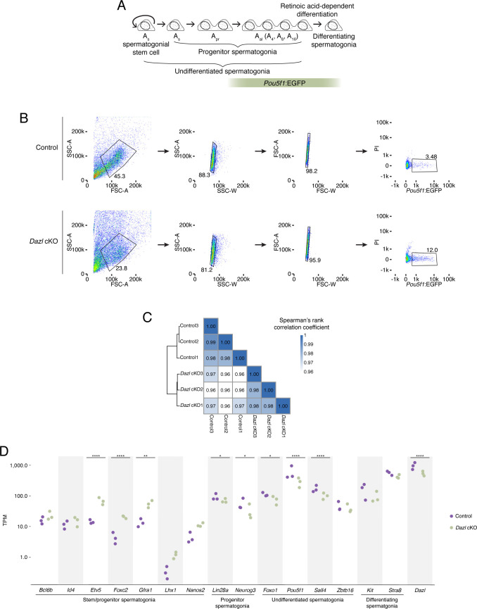
Figure 1—figure supplement 3. Characterization of Pou5f1:EGFP-positive spermatogonia in Dazl cKO.

Figure 1—figure supplement 3.

(A) Schematic of spermatogenesis from undifferentiated spermatogonia through differentiating spermatogonia, with expression of Pou5f1:EGFP. (B) Schematic depicting the sorting strategy used to isolate Pou5f1:EGFP-positive spermatogonia from control and Dazl cKO adult testes. The GFP+ population (right panel) was collected for RNA-seq. (C) Heatmap of sample-to-sample distances among RNA-seq datasets of sorted Pou5f1:EGFP-positive spermatogonia from control and Dazl cKO testes. Three biological replicates were analyzed for each genotype. (D) Differential gene expression analysis of RNA-seq analysis of Pou5f1:EGFP-positive spermatogonia from adult testes, highlighting genes associated with spermatogonial stem cells and early progenitors (‘stem/progenitor spermatogonia’), progenitor spermatogonia, undifferentiated spermatogonia (i.e., spermatogonial stem cells as well as early and late progenitors), and differentiating spermatogonia. * adjusted p<0.05, ** adjusted p<0.01, *** adjusted p<0.001, **** adjusted p<0.0001.