. 2020 Jul 20;9:e56523. doi: 10.7554/eLife.56523

Figure 2. Identification of DAZL-bound transcripts in undifferentiated spermatogonia via iCLIP reveals DAZL’s regulation of spermatogonial factors.

(A) Schematic of the synchronization of spermatogenesis to obtain undifferentiated spermatogonia via the 2S method for iCLIP (green arrows) and via the 3S method for RNA-seq (purple arrows). WIN 18,446 was used to synchronize spermatogenesis by blocking spermatogonial differentiation, and samples were histologically staged to verify successful synchronization. For 3S samples, germ cells were sorted from synchronized testes. (B) Genomic distribution of DAZL iCLIP peaks identified in three biological replicates (TPM ≥1; FDR < 0.05). (C) Venn diagram showing overlap of DAZL iCLIP peaks in expressed 3' UTRs (TPM ≥1) among three biological replicates. Replicated peaks (i.e., present in at least two of three replicates) were identified. After merging replicated peaks that fell on consecutive nucleotides, 11,882 DAZL binding sites (present in at least two of three replicates; highlighted in blue) were identified. These binding sites correspond to 2,633 genes, which are designated as the DAZL-bound genes. (D) Enrichment of factors that regulate the development and differentiation of undifferentiated spermatogonia in DAZL targets compared with all genes expressed in undifferentiated spermatogonia (one-tailed hypergeometric test). Number of genes (n) in each group designated in parentheses in labels along x-axis.****, p<0.0001. (E) DAZL iCLIP, IgG iCLIP, and 3S RNA-seq gene tracks showing exemplary DAZL-bound genes that are required for spermatogonial proliferation and expansion (Lin28a and Sox3) or differentiation (Kit, Foxo1, Rarg, and Sall4). Each iCLIP track represents the crosslinked sites from the sum of unique reads from three biological replicates. The RNA-seq track represents the sum of two biological replicates. The scale of each gene track is marked on the left. 3' UTRs are in light blue.

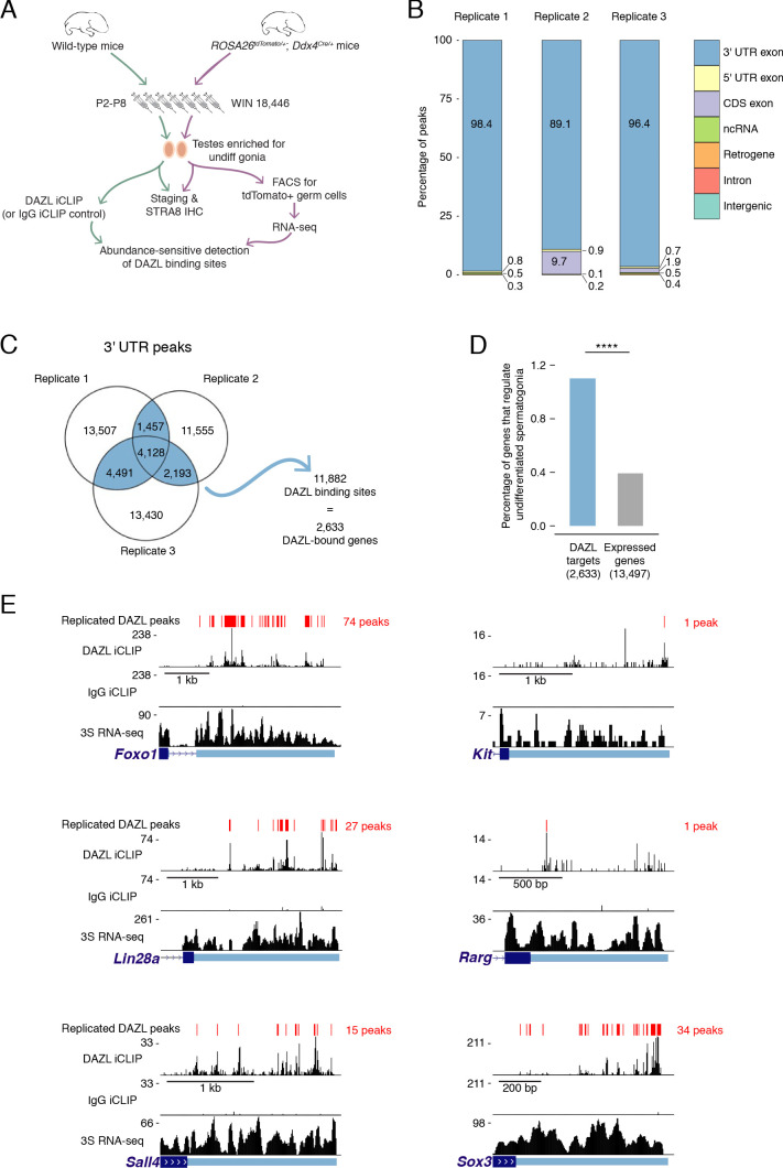
Figure 2—source data 1. Genes that regulate development and differentiation in undifferentiated spermatogonia.
Figure 2—source data 2. Replicated DAZL iCLIP peaks and transcript expression levels in 3S undifferentiated spermatogonia.
Figure 2—source data 3. Comparison of 3S undifferentiated spermatogonia to previously published spermatogonial datasets.

Figure 2.

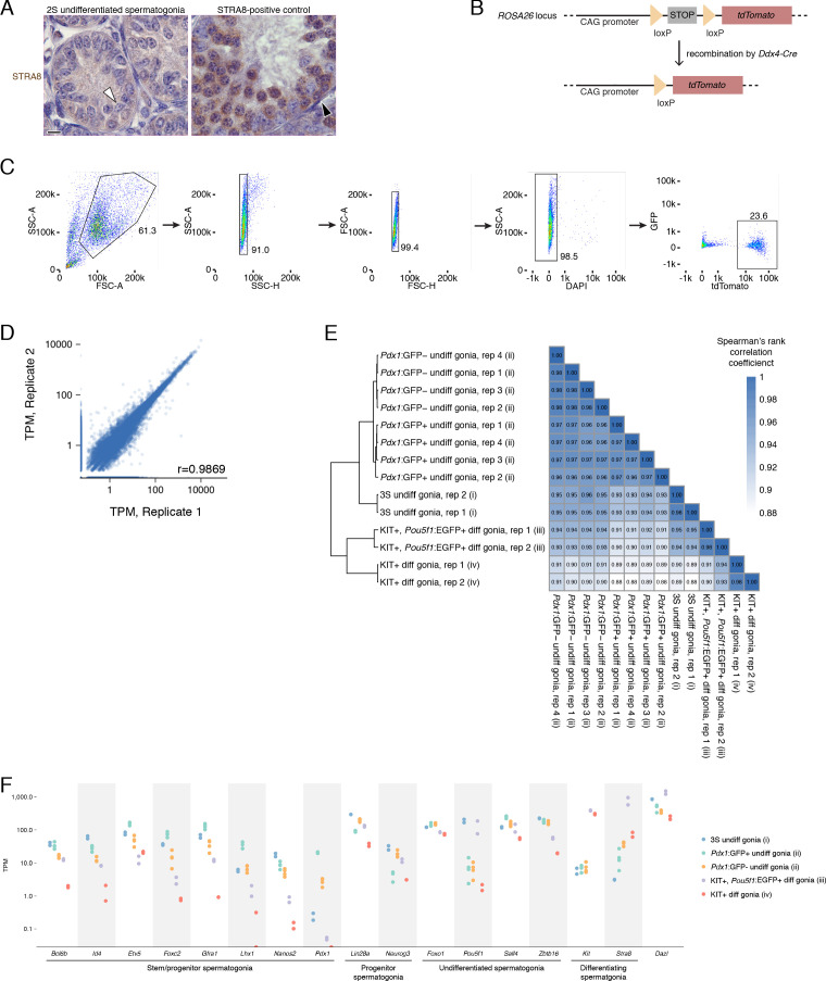
Figure 2—figure supplement 1. Isolation of undifferentiated spermatogonia via the 2S (synchronization and staging) and 3S (synchronization, staging, and sorting) strategies.

Figure 2—figure supplement 1.

(A) The absence of STRA8 expression in WIN 18,446-synchronized testes immunohistologically confirms the accumulation of undifferentiated spermatogonia. White arrowhead highlights a STRA8-negative type A spermatogonium. Testes synchronized for preleptotene spermatocytes were used as a positive control for STRA8 immunohistology. Black arrowhead highlights a STRA8-positive differentiating spermatogonium; the majority of the STRA8-positive cells in this section are preleptotene spermatocytes. (B) Schematic of the Cre reporter tdTomato. Recombination of the tdTomato allele via the Ddx4Cre allele removes the floxed stop codon and activates expression of tdTomato protein specifically in germ cells. (C) Gating strategy for sorting undifferentiated spermatogonia after synchronization. Undifferentiated spermatogonia were sorted from 2S testes enriched for undifferentiated spermatogonia. The DAPI-negative population represents the live cell fraction, and the tdTomato-positive population represents the undifferentiated spermatogonia that were collected for RNA-seq. (D) Correlation between biological replicates of TPM expression values for protein-coding genes, noncoding RNA (excluding rRNA), and retrogenes with TPM ≥1. Pearson’s correlation coefficient shown. (E) Heatmap of sample-to-sample distances between RNA-seq datasets from (i) our 3S undifferentiated spermatogonia; (ii) Pdx1:GFP-positive undifferentiated spermatogonia and Pdx1:GFP-negative undifferentiated spermatogonia from 6 to 8 week old adult testes La et al., 2018a; (iii) KIT-positive, Pou5f1:EGFP-positive differentiating spermatogonia from P7 testes Kubo et al., 2015; (iv) KIT-positive differentiating spermatogonia from P7 testes (Maezawa et al., 2017). RNA-seq data for protein-coding genes were quantified, normalized and transformed via DESeq prior to calculating sample distances. (F) Expression of markers genes for spermatogonial stem cells and early progenitors (‘stem/progenitor spermatogonia’), progenitor spermatogonia, undifferentiated spermatogonia (i.e., spermatogonial stem cells as well as early and late progenitors), and differentiating spermatogonia in the RNA-seq datasets analyzed in E.
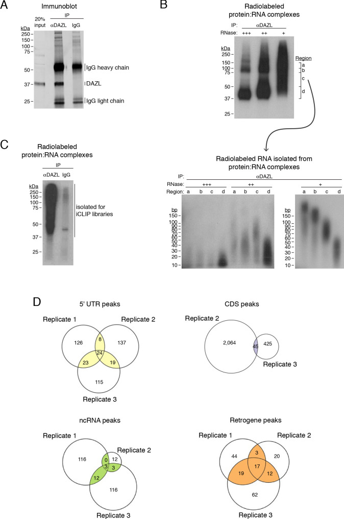
Figure 2—figure supplement 2. DAZL iCLIP.

Figure 2—figure supplement 2.

(A) DAZL immunoblot from P20 testes. Immunoprecipitation (IP) and Western blotting identifies DAZL at ~37 kDa. (B) Radiolabeled DAZL:RNA complexes from P20 testes (upper blot) and radiolabeled RNA isolated from these DAZL:RNA complexes (lower blot). Prior to radiolabeling, DAZL:RNA complexes were subjected to digestion by RNAse I at high (+++), medium (++), and low (+) concentrations. High RNAse concentration identifies DAZL:RNA complexes just above 37 kDa, where DAZL protein alone is found, and at ~75 kDa, which is consistent with DAZL:RNA complexes containing two DAZL proteins bound to a single RNA fragment. To obtain RNA of a sufficient length for preparing cDNA libraries, the low concentration of RNAse I was used to prepare iCLIP libraries. (C) Radiolabeled DAZL:RNA complexes from postnatal testes synchronized for undifferentiated spermatogonia. One of three biological replicates used to prepare iCLIP libraries is shown. (D) Venn diagram showing overlap of DAZL iCLIP peaks in expressed transcripts (TPM ≥1) from each type of genomic region, other than 3' UTRs, among three biological replicates.