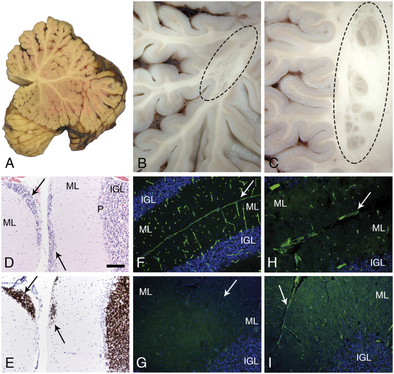
FIGURE 4.
Cerebellar pathology. A mid-sagittal section of the cerebellum showed normal gross morphology (A). In contrast, several examples of subcortical, heterotopia was observed in the hemispheres (B, C; oval dashed lines). Microscopic evaluation revealed sites of failed external granule cell migration (arrows in D, H&E and E, neuN; IGL, P, ML). Immunofluorescence staining of cryosections revealed merosin at the pial surface basement membrane and perivascular basement membranes of control cerebellum (F); merosin was absent from the patient’s cerebellum (G; arrow marks the position of a sulcus). Other laminins were present at the patient’s pial surface and perivascular basement membranes (H, polyclonal laminin, α1 subunit on laminin-111; I, laminin a5). Arrows in H and I mark the position of sulci. The pial surface basement membrane was interrupted at some sites (H). The scale bar in panel D is 100 µm for panels C–I. H&E, hematoxylin and eosin; IGL, internal granule cell layer; ML, molecular layer; P, Purkinje cell layer.

