Fig. 1.
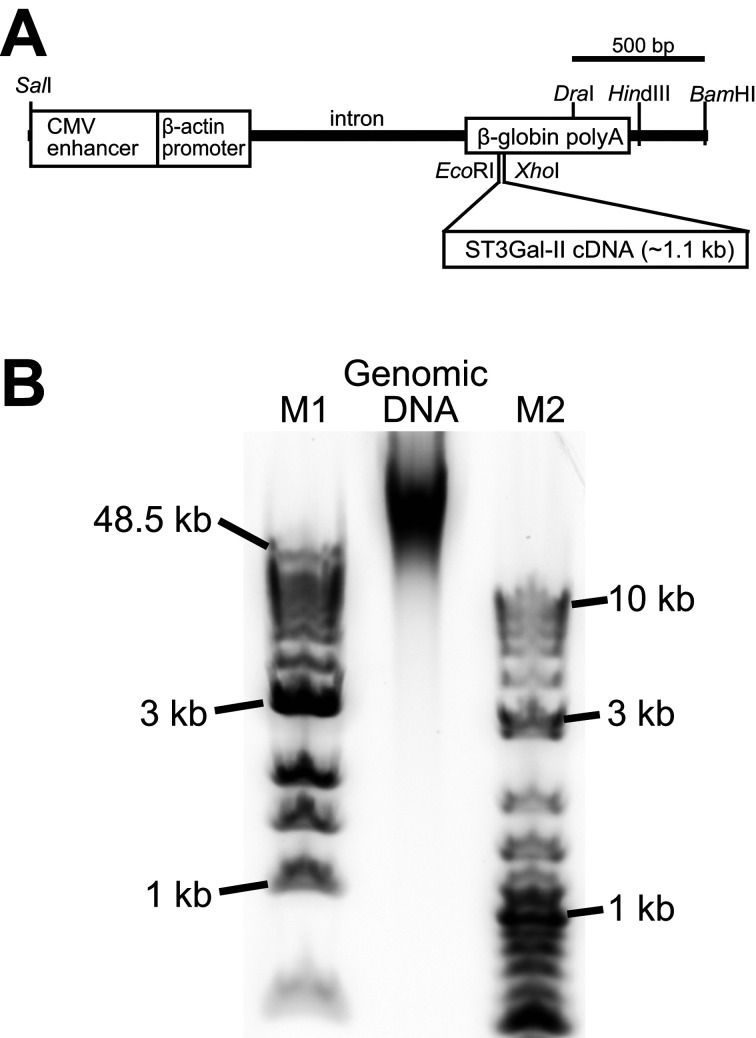
Transgene structure and DNA size of a genomic DNA sample. (A) Transgene structure used for production of 4C30 mice (accession number: LC495729) [39]. The transgene (3,508 bp) was a SalI-BamHI fragment derived from the pCAGGS vector containing cytomegalovirus enhancer, chicken β-actin promoter, and rabbit β-globin polyadenylation signal sequences [29]. The cDNA of mouse ST3 β-galactoside α-2,3-sialyltransferase 2 (ST3GalII, a.k.a., St3gal2 and Siat5) [22], a mouse sialyltransferase, was inserted into EcoRI-XhoI sites in the β-globin polyadenylation signal sequence. (B) DNA size distribution of a genomic DNA extracted from a 4C30 mouse kidney. Note that most genomic DNAs in this sample were longer than 50 kbp. The genomic DNA sample (~0.4 µg) and molecular markers (~0.5 µg) were separated with 1% agarose gel electrophoresis (E-gel EX 1%, Thermo Fisher Scientific). The electropherogram was recorded using a laser scanner (FX-Pro, Bio-rad). M1: 1 kb extended DNA ladder (NEB), M2: 2-log DNA ladder (NEB).

