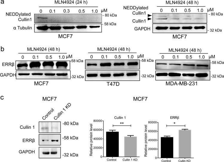
Fig. 2. ERRβ expression is regulated by NEDDylation and the SCF complex complex in breast cancer.
a MCF7 cells were treated with varying concentrations of MLN4924 (0, 0.1, 0.3, 0.5 and 1.0 µM) for 24 h prior to western blot analysis of NEDDylated Cullin1. The NEDD8-modified Cullin1 was determined by probing the whole western blot membrane with an anti-NEDD8 antibody. The characteristic predominant band (just below 80kD) correspond to the NEDD8-modified Cullin1, were shown. Tubulin was used as a loading control (left panel). MCF7 cells were treated with varying concentrations of MLN4924 (0, 0.1, 0.3, 0.5 and 1.0 µM) for 48 h prior to western blot analysis of hyper-NEDDylated and hypo-NEDDylated Cullin1. GAPDH was used as a loading control (right panel). b MCF7, T47D and MDA-MB-231 cells were treated with varying concentrations of MLN4924 (0, 0.1, 0.5 and 1.0 µM) for 48 h prior to western blot analysis of ERRβ. GAPDH was used as a loading control. (N = 3). c Expression levels of ERRβ and Cullin1 in MCF-7 cells were analysed by western blotting after transfection with siRNA targeting Cullin1 or non-targeting control siRNA. Western blots are representative of three independent experiments. The ratio to GAPDH expression was calculated for the relative ERRβ and Cullin1 expression levels. The relative expression levels (right panels) are shown. Data represent means ± SD. (n = 3; t tests). Significant *P < 0.05; **P < 0.01.

