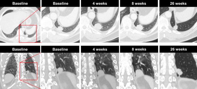
Fig. 1.
A case of pseudoprogression in a 49-year-old man with NSCLC who was receiving immune checkpoint inhibitor therapy. Coronary CT imaging obtained before therapy demonstrates a 20 mm metastasis in the left lower lobe and 4 weeks after treatment initiation, the tumor size increased from 20 to 29 mm. Therapy was continued, and 4 weeks thereafter, the tumour size was reduced by 4 mm to 25 mm. Treatment was continued and 26 weeks after treatment initiation, the tumor continued to reduce in size, measuring 14 mm. No further metastases were visible at any time point. Upper row: axial view, lower row: cornonal view

