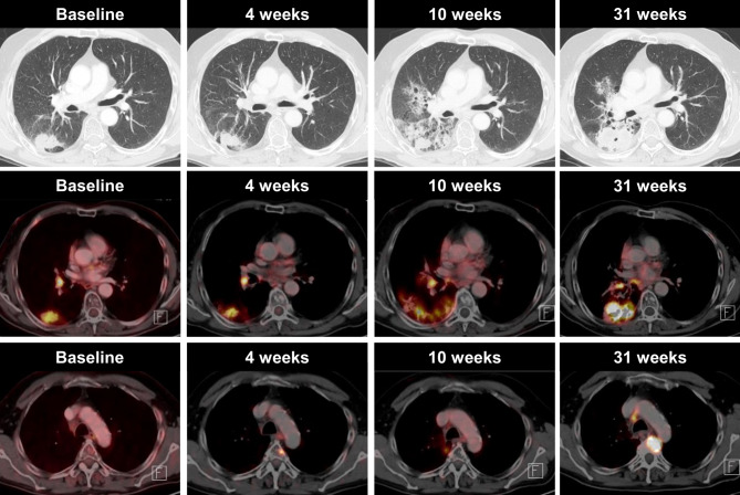
Fig. 2.
A case of pseudoprogression in a 52-year-old man with NSLCL, who was receiving immune checkpoint inhibitor therapy. Axial CT and 18F‑FDG-PET/CT imaging obtained before the start of treatment show a metabolically active tumor in the right lower lobe and an ipsilateral hilar lymph node metastasis. Four weeks after treatment initiation, the tumor slightly decreased in size and showed reduced 18F‑FDG uptake. However, we observed a newly developed, metabolically active metastasis in the 4th thoracic vertebral body measuring <1.0 cm. Treatment was continued and PET-CT performed 10 weeks after the start of treatment showed no increased 18F‑FDG uptake in the 4th thoracic vertebral body. The 18F‑FDG uptake in the primary tumour in the right lower lobe further decreased. The newly developed, metabolically active, ground-glass opacities were radiation-associated. Thirty-one weeks after the start of treatment, 18F‑FDG PET-CT evidenced disease progression in both the primary tumour and the metastasis in the 4th thoracic vertebral body. Upper row: CT in lung window, middle row: axial fused PET/CT image lower row: axial fused PET/CT image

