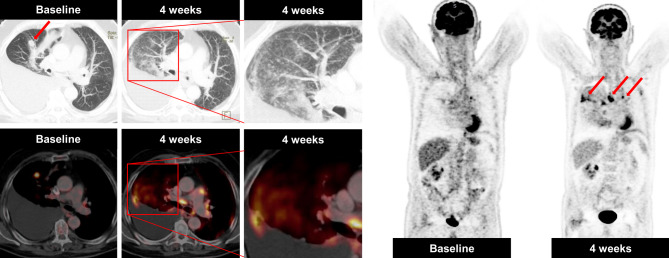
Fig. 3.
A case of a sarcoid-like reaction in a 76-year-old male NSCLC patient, who was receiving immune checkpoint inhibitor therapy. Axial CT in lung window and fused 18F‑FDG-PET/CT imaging obtained before treatment initiation showed a metabolically active metastasis in the middle lobe and a right pleural effusion. Four weeks after treatment initiation, multiple intrapulmonary micronodules in a perilymphatic distribution were detectable in both lungs, predominately right ≫ left. In addition, enlarged metabolically active hilar/mediastinal lymph nodes were visible (arrows), consistent with a sarcoid-like reaction. The disappearance of the metastasis in the middle lobe was suggestive of treatment response

