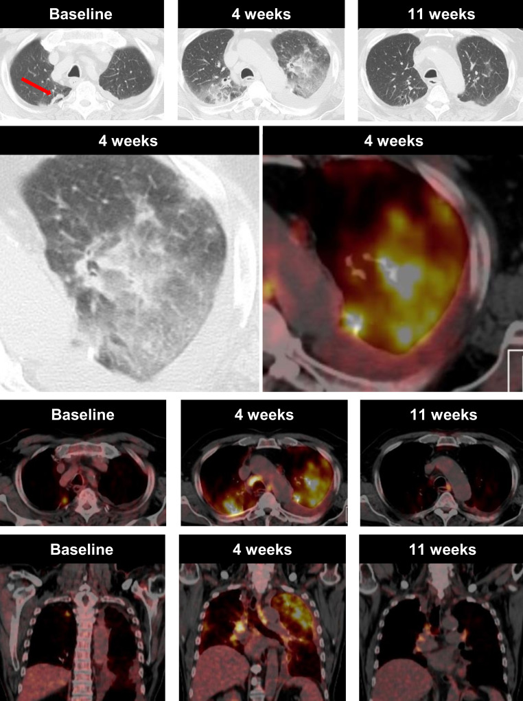
Fig. 4.
A case of pneumonitis in a 75-year-old male NSLCL patient, who was receiving immune checkpoint inhibitor therapy. Axial and coronal CT in lung window and fused 18F‑FDG-PET/CT imaging obtained before treatment initiation show a metabolically active metastasis in the right upper lobe, as well as five cerebral metastases (not shown). Four weeks after treatment initiation, the pulmonary, as well as the cerebral metastasis (not shown), showed a significant tumour shrinkage. In contrast, new, patchy, highly metabolically active ground-glass opacities in both lungs (upper lobes > lower lobes), as well as metabolically active enlarged mediastinal lymph nodes, were visible. On the day of the examination the treatment was discontinued and the patient was started on cortisone. After 7 weeks the pulmonary changes resolved almost completely while slightly enlarged metabolically active mediastinal lymph nodes remained. Upper three rows: axial view; lower row: coronal view

