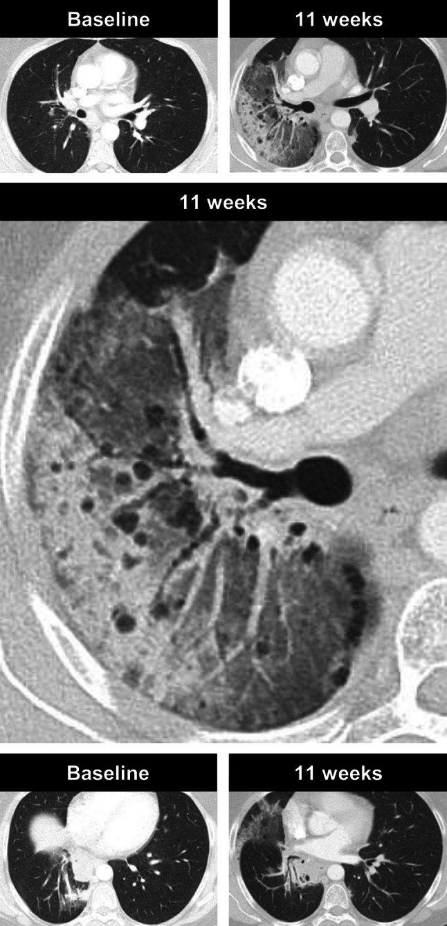
Fig. 5.

A case of pneumonitis in a 52-year-old female NSLCL patient, who was receiving immune checkpoint inhibitor therapy. Axial CT images in lung window obtained before treatment initiation show no evidence of malignant disease and 11 weeks after treatment initiation, she developed considerable ground-glass opacities and consolidations in the right upper and middle lobe without pleural effusion or signs of lymphangiosis carcinomatosa. Treatment was paused and the patient was started on cortisone. Thereafter, her general condition improved rapidly
