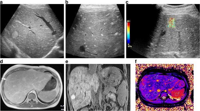
Fig. 10.
Ultrasound elastography and MRI in Fontan-associated liver disease in a 16-year-old girl with fibrosis, cirrhosis and nodular liver lesions. a Sagittal US imaging reveals a heterogeneous parenchyma and a large nodular lesion of 2 cm in diameter laterally in the right liver lobe. b, c Transverse linear high-frequency US probe reveals multiple small hyperechoic lesions (b), and transverse shear-wave elastography shows increased liver stiffness (c). d Axial T1-W gradient echo MR image post gadolinium contrast injection reveals an enlarged congestive liver with irregular enhancement of the nodule (arrow). e Coronal T1 gradient echo image shows strong enhancement of the nodule with gadoxetic acid in late hepatobiliary phase, interpreted as a focal-nodular-hyperplasia-type lesion. f MR native T1 mapping in an axial midsection plane, with regions of interest drawn in the liver and spleen, shows irregular increased T1 times in the periphery

