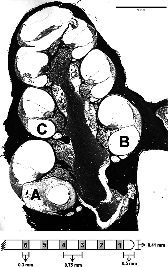
Fig. 1.
Mid-modiolar section of a guinea pig cochlea (micrograph) illustrates the implant location in the scala tympani (profile A) and the profiles of interest inside the cochlea that were used for SGN density and tissue density analysis (profiles A, B, and C in bold). Also shown is a schematic of an eight-electrode scala tympani cochlear implant, (below the micrograph); only the first 6 potentially intracochlear electrodes are shown

