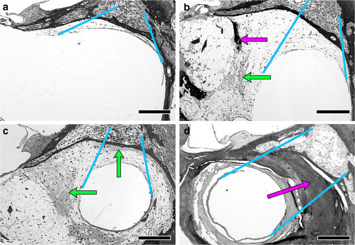
Fig. 2.
Variability of intrascalar tissue found in implanted guinea pigs. a Minimal tissue. b More extensive tissue with patches of bone (dark stain and laminated) and cartilage (lighter and unlaminated). c Similar to b, but with cartilage between the implant and the spiral ganglion. d Implant completely surrounded by bone that fills most of the scala. Blue lines illustrate the criteria used to delineate the regions of interest for scoring the intrascalar tissue (the tissue potentially between the SGN cell bodies in Rosenthal’s canal and the implant), connecting the edges of the area occupied by the SGN to the edges of the open space in scala tympani. Arrows indicate patches of cartilage (green) and bone (magenta) in the intrascalar tissue. Bar = 200 μm. Panel a is an example of a cochlea classified as Low intrascalar tissue, b is an example of Medium intrascalar tissue, and c and d are examples of High intrascalar tissue. See text for further details

