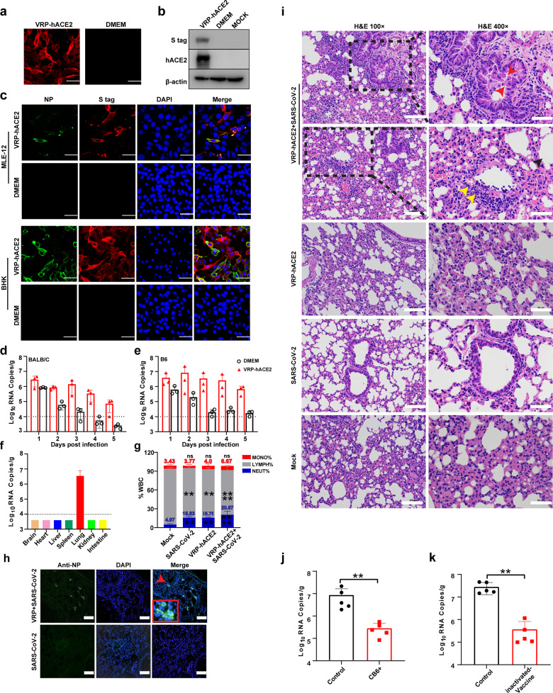
Fig. 1. Development of mouse model for SARS-CoV-2 infection.
a, b Expression of hACE2 was analyzed using IFA (a) and western blot assay (b) in MLE-12 cells transduced with VEEV-VRP-hACE2. c Transduction with VEEV-VRP-hACE2 converted nonpermissive MLE-12 cells and BHK-21 cells into SARS-CoV-2 permissive cells. d, e Viral RNA loads of lung tissues in VEEV-VRP-hACE2-transduced BALB/c (d) and C57BL/6 mice (e) after infection with SARS-CoV-2. f Viral RNA loads of different tissues in the BABL/c mice transduced with VEEV-VRP-hACE2 followed by SARS-CoV-2 infection. g White blood cells analysis in the peripheral blood collected from each group of BALB/c mice. h SARS-CoV-2 antigen detection in the lung tissues of BALB/c mice transduced with VEEV-VRP-hACE2 and infected with SARS-CoV-2 using anti-NP antibody. i H&E staining of lung samples from infected BALB/c mice at 4 dpi. j Viral RNA loads of lungs in BALB/c mice inoculated with CB6 at 2 days post challenge with SARS-CoV-2. k Viral RNA loads of lungs in BALB/c mice twice immunized with inactivated SARS-CoV-2 vaccines at 2 days post challenge with SARS-CoV-2. Data were expressed as means ± SD, Student’s t test was used to analyze the differences between two groups. **P < 0.01, ***P < 0.001. Scale bars, 50 μm (a, c), 100 μm (h, i left), 50 μm (i right).

