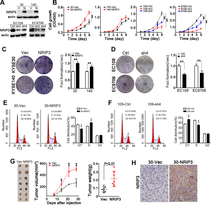
Fig. 2. NRIP3 is functionally critical for ESCC tumor cell growth.
a Western blotting analysis of NRIP3 expression in the indicated cell lines (V vector; N NRIP3). β-actin was used as a loading control. b The cell growth rate was evaluated in NRIP3-overexpressing cells (KYSE30 and KYSE140) and NRIP3-KD cells (EC109 and EC9706) (*P < 0.05; **P < 0.01). c Representative images and summary of foci formation in NRIP3-overexpressing cells (KYSE30 and KYSE140) (**P < 0.01). d Representative pictures and summary of foci formation in NRIP3-KD cells (EC109 and EC9706). Cell cycle was analyzed by flow cytometry in NRIP3-overexpressing cells (e) and NRIP3-KD (f) cells (left: representative images; right: summary). g The image of xenografts formed in the nude mice (left), tumor growth curve (middle) and tumor weight (right) are summarized (**P < 0.01). h Representative IHC staining images comparing NRIP3 expression between 30-Vec and 30-NRIP3 xenografts (original magnification: ×200).

