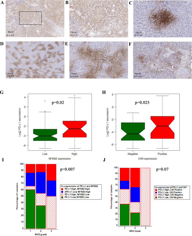
Figure 2.
Representative images (× 20) from IHC staining of high cytoplasmic expression of NFKB2 (A,B) in a meningioma grade II. The CA9 IHC staining shows presence of regional, membranous and cytoplasmic in the tumor; distant from vascular channels in WHO grade II meningioma (C,D) and also in per necrotic areas of WHO grade III (E,F). The Box plot shows significant higher percentage of PD-L1 positive cells in meningioma with high NFKB2 expression (G). The same analysis in primary meningiomas indicates higher percentage of PD-L1 in positive CA9 meningioma cases (H). Chi-squared analysis demonstrated the positive association of WHO grade and co-expression of NFKB2 and PD-L1 in the meningioma cohort (I). The same analysis for WHO grade and co-expression of CA9 and PD-L1 (J). (R v3.3.1, MedCalc).

