Figure 2.

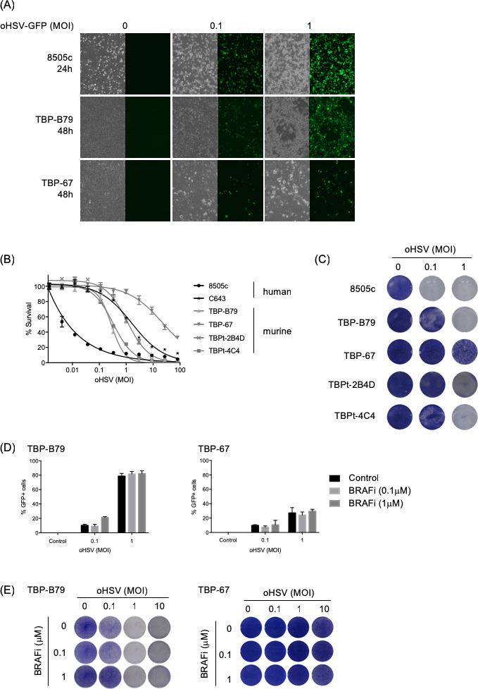
In vitro effects of oHSV on human and murine thyroid cancer cell lines. (A) Human (8505 c) and murine (TBP-B79 and TBP-67) thyroid cancer cell lines were infected with oHSV-GFP at various MOIs and cells were imaged for virus replication (visualized by GFP signal) at indicated time points post-infection. Corresponding bright-field images show cell morphology changes following virus infection. (B) Human (8505 c and C643) and murine (TBP-B79, TBP-67, TBPt-2B4D and TBPt-4C4) thyroid cancer cell lines were infected with 1:3 dilutions of oHSV starting at MOI 100 or left uninfected as controls and cell survival was measured 72 hours later using CellTitreGlo assay. (C) Human (8505 c) and murine (TBP-B79, TBP-67, TBPt-2B4D and TBPt-4C4) thyroid cancer cell lines were infected with oHSV (MOI 0.1 or 1) or left uninfected and cells were fixed and stained with crystal violet 48 hours later. (D) Murine thyroid cancer cell lines (TBP-B79 and TBP-67) were infected with oHSV-GFP (MOI 0.1 or 1) or left uninfected, and subsequently treated with BRAF inhibitor (BRAFi) (0.1 or 1 µM). Cells were analyzed for GFP positivity (virus infection) using flow cytometry 48 hours post-treatment. (E) Murine thyroid cancer cell lines (TBP-B79 and TBP-67) were infected with oHSV at various MOI and treated with BRAFi at indicated concentrations. Cells were fixed and stained with crystal violet 48 hours later. Each experiment was performed a minimum of two times with similar results. BRAFi, BRAF inhibitor; GFP, green fluorescent protein; oHSV, oncolytic herpes simplex virus.
