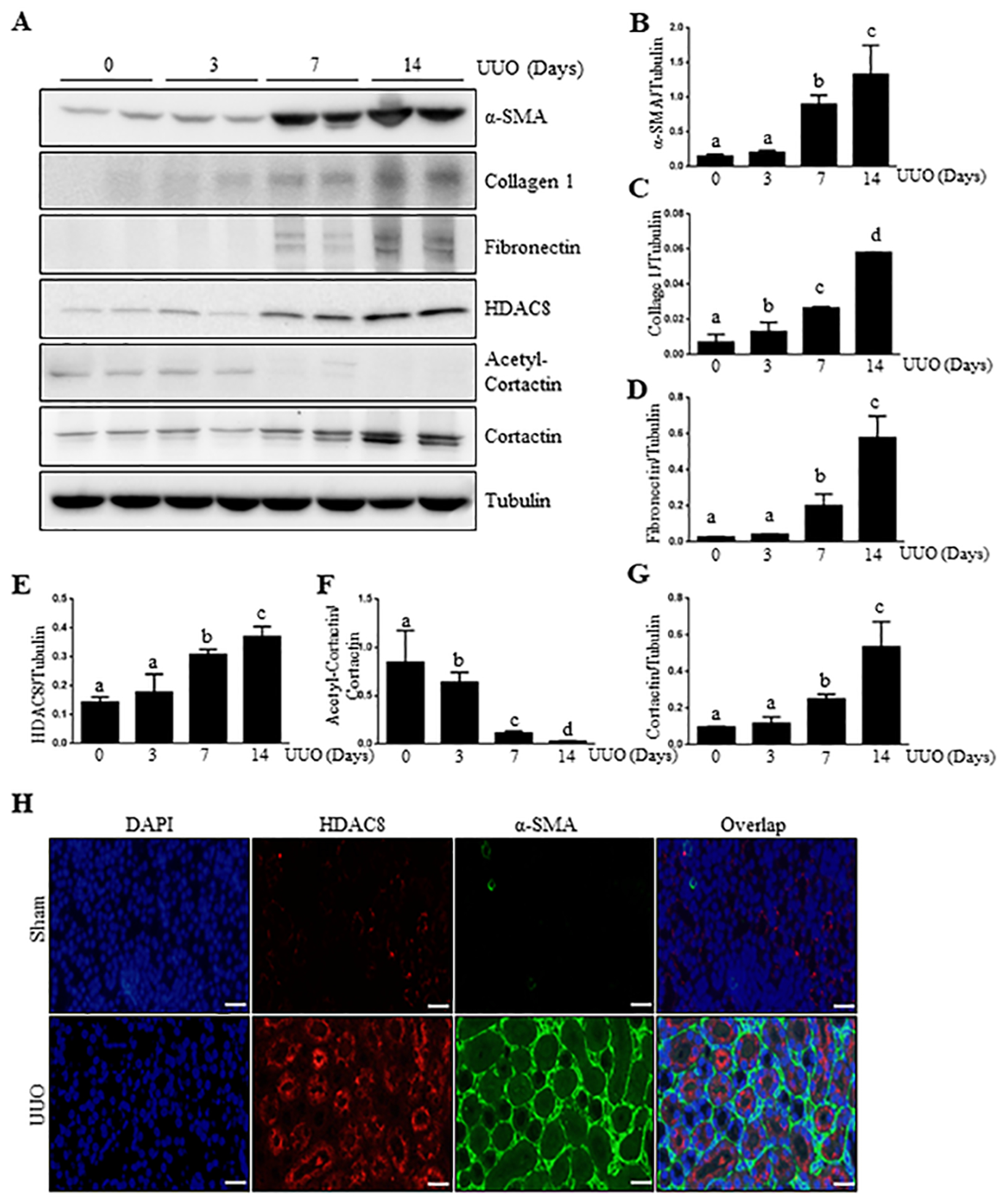
Figure 1. UUO induces HDAC8 expression and development of interstitial fibrosis over time.

(A) The kidneys were harvested at 0, 3, 7, 14 days after surgery with left ureteral ligation and then homogenized. Kidney tissue lysates were subjected to immunoblot analysis with antibodies against α-SMA, Collagen1, Fibronectin, HDAC8 and tubulin, respectively. Expression levels of α-SMA, Collagen1, Fibronectin, HDAC8 and Tubulin were performed were quantified by densitometry and the levels of α-SMA (B), Collagen1 (C), Fibronectin (D), HDAC8 (E) and Cortactin (G) were normalized with Tubulin. Acetyl-Cortactin (F) was normalized with Cortactin. Values are the means ± S.D (n = 6). Means with different superscript letters (a-d) are significantly different from one another (P<0.05). (H) Photomicrographs illustrating HDAC8 (red), α-SMA (green) and nuclei (blue) immunofluorescent staining of kidney tissue after UUO treatments (200X). Scale bar: 50 μM.
