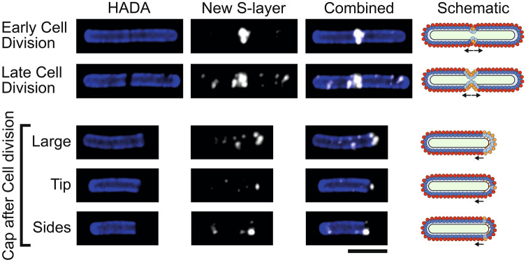
Figure 3.
S-layer formation during cell division. Airyscan confocal images of C. difficile 630 cells during and immediately after cell division with HADA labelled peptidoglycan cell wall (blue) and new surface SlpAR20291 immunolabeled with Cy5 (white). Large, dark areas lacking HADA staining mark peptidoglycan synthesis at the septum between cells or a newly produced cell pole. Scale bar indicates 3 µm. On the right-hand side of each row is a schematic diagram illustrating the position of new surface SlpAR20291 (HMW-SLP, spotted light blue and LMW-SLP, spotted orange) as detected in the corresponding microscopy images against the position of endogenous surface SlpA630 (HMW-SLP, dark blue and LMW-SLP, red). The position of newly synthesized cell wall is displayed in brown/white stripes.

