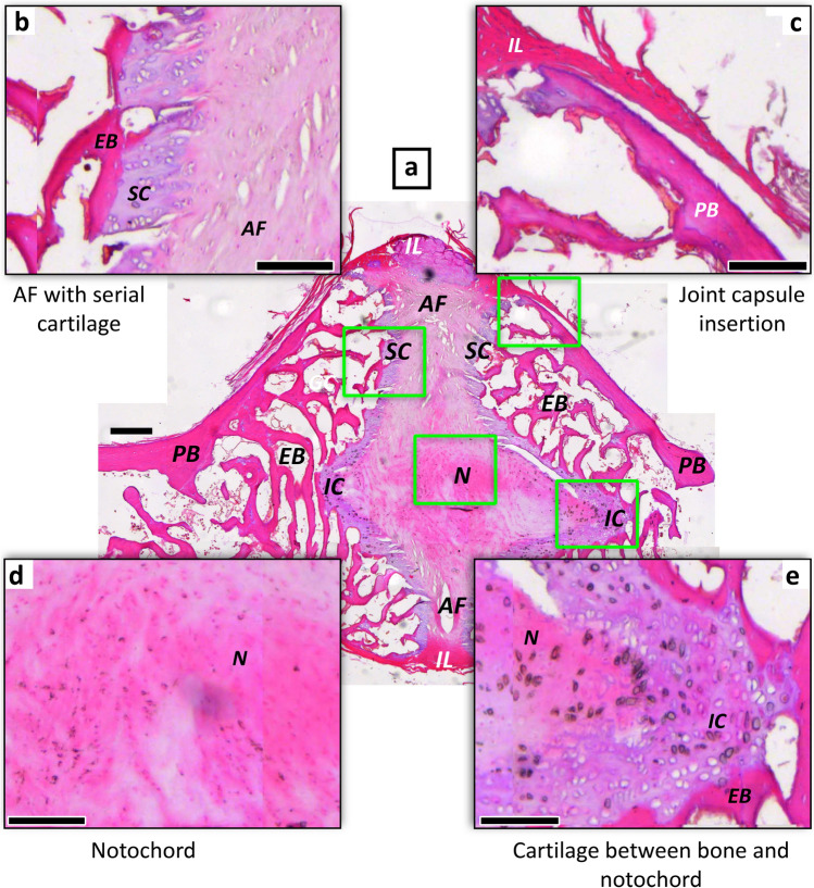
Figure 1.
Histology of the plesiomorphic amniote joint with persisting notochord of Sphenodon punctatus NHMW 8108. The intervertebral tissues and the two articulating mid-dorsal centra illustrate the relationship between mineralised (fossilisable) tissues and soft tissues. (a) Oblique sagittal microtome section stained with hematoxylin. Note that the notochord appears discontinuous in the image because of the suboptimal plane of section. (b) Enlargement of annulus fibrosus insertion into endochondral bone of articular surface via serial cartilage. (c) Enlargement of interspinal ligament of the joint capsule inserting into periosteal bone of the centrum peripheral surface. (d) Enlargement of notochordal tissue in intervertebral space. (e) Enlargement of contact between notochordal tissue and endochondral bone of articular surface of centrum with intervening irregular cartilage. Scale bar in (a) represents 100 µm, and scale bars in (b–e) represent 40 µm. AF annulus fibrosus, EB endochondral bone, IC irregular cartilage, IL intervertebral ligament, N notochordal tissue, PB periosteal bone, SC serial cartilage.

