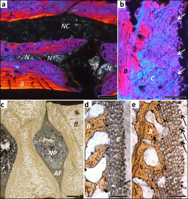
Figure 3.
Histology of mesosaur, ichthyosaur and dinosaur intervertebral spaces, all fossil ground sections. (a) Stereosternum tumidum IGPB R 622, sagittal section of two articulated dorsal vertebral centra with intervertebral space, showing the notochordal amphicoelous shape and the persisting notochord. Image is in cross-polarised light with lambda filter. (b) Hadrosauridae indet. UALVP 59650. Close up of the articular surface showing obliquely arranged mineralised fibres in between poorly defined files of chondrocyte lacunae (arrows). Image is in cross-polarised light with lambda filter. (c) Stenopterygius sp. IGPB R 661 sagittal section of two articulated centra showing the amphicoelous shape. Note the differentiation of the content of the intervertebral space into a coarse into a fine fraction, probably representing the nucleus pulposus (NP) and the annulus fibrosus (AF). (d) Enlargement of the concave part of the articular surface, showing a thin layer of irregularly arranged chondrocyte lacunae, underlying the nucleus pulposus (NP). (e) Enlargement of the convex part of the articular surface, showing obliquely arranged files of chondrocyte lacunae, representing the insertion of the annulus fibrosus (AF) (arrows). Scale bar in (a) represents 500 µm, scale bar in (b) represents 100 µm, scale bar in (c) represents 2 mm, scale bars in (d, e) represent 100 µm. AF annulus fibrosus, B bone tissue, C cartilage, N notochord, NC neural canal, NP nucleus pulposus.

