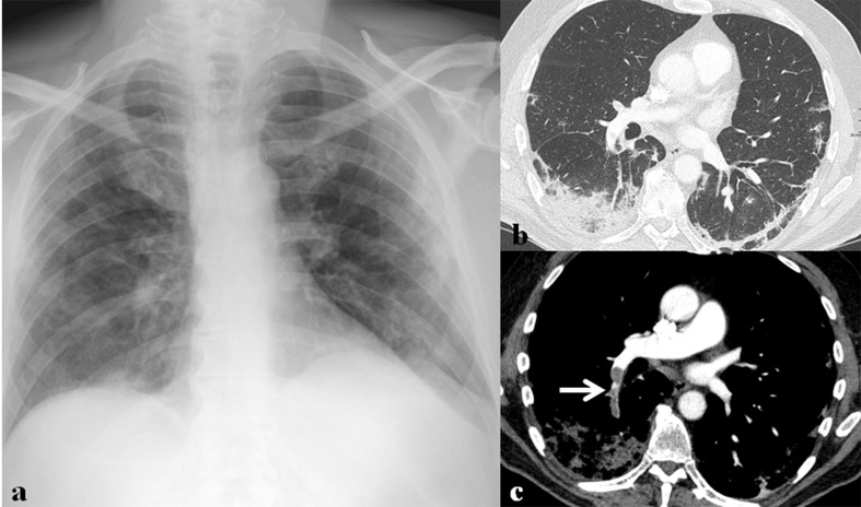
Figure 14.
A 71-year-old male patient with SARS-CoV-2 infection. (a) Initial chest radiograph shows bilateral peripheral ground-glass opacities (GGOs) and multifocal patchy consolidation in both lungs. (b) Lung window image of contrast-enhanced chest CT scan obtained on the same day with (a) shows consolidation and patchy GGOs in the subpleural areas of both lower lobes. (c) Mediastinal window image of contrast-enhanced chest CT scan obtained on the same day with (a) shows thromboembolism (arrow) involving right lower lobar pulmonary artery.

