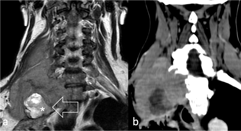
Figure 10.
50-year-old male with spindle cell/sclerosing RMS located in the inter scalene triangle (arrow) visualised (a) at T2W and (b) at contrast-enhanced CT, involving right supraclavicular nodes, I° rib and the apex of the lung. The hyperintense lesion within the mass (asterisk) was revealed to be a neurofibroma. T2 W, T2 weighted,

