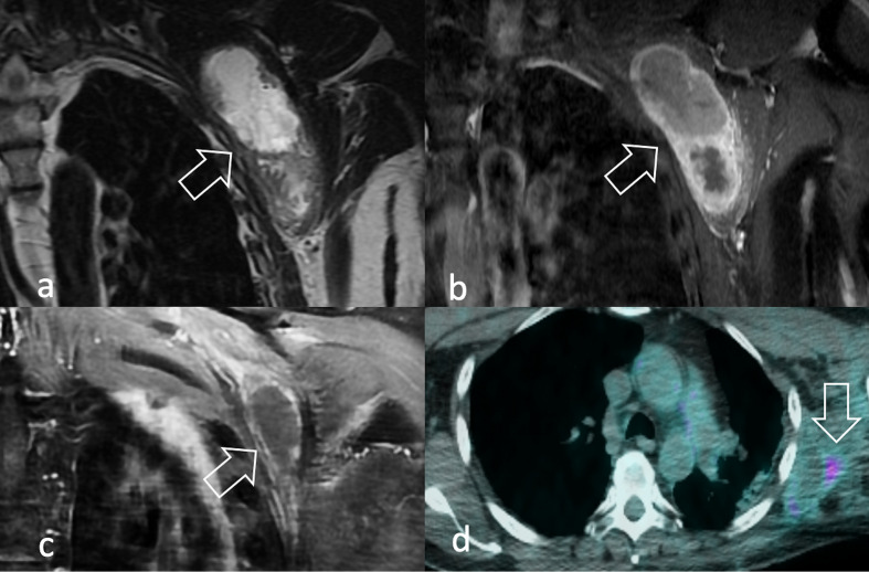
Figure 11.
A 59-year-old male presenting with a lump in his left axilla. MRI showed on coronal T2W (a) and T1W after contrast-enhancement (b) a vascularised, extensively necrotic mass in the left axilla (arrow). Diagnosis of pleomorphic RMS was assigned after biopsy. After (c) partial response after chemotherapy (arrow), the lesion was surgically removed with radical intent and adjuvant radiation therapy was delivered. 18F-FDG PET/CT performed 1 year later showed (d) minor uptake, consistent with chronic inflammation (arrow). 18F-FDG, 18F-fludeoxyglucose; PET, positron emision tomography; RMS, rhabdomyosarcomas; T1W, T1 weighted; T2W, T2 weighted.

