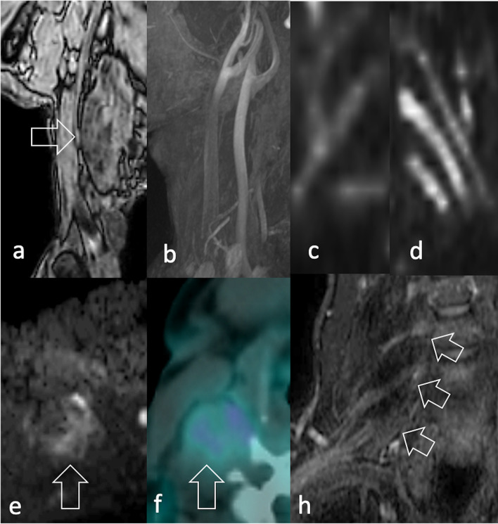
Figure 12.
Same patient in Figure 2e–f. A 52-year-old male with pain in his right upper extremity during exercise. (a) T1W image MRI shows an 8 cm diameter mass lying on the scaleni muscles, without (b) arterial involvement (c) DWI neurography displays the involvement of the brachial plexus compared to (d) the contralateral plexus. (e) Peripheral signal restriction (arrow) (f) mild radiopharmaceutical uptake (arrow) were identified at DWI and 18F-FDG PET/CT, respectively. Biopsy revealed spindle cell/sclerosing RMS. 18F-FDG, 18F-fludeoxyglucose; DWI, diffusion-weighted imaging; PET, positron emision tomography; RMS, rhabdomyosarcomas; T1W, T1 weighted.

