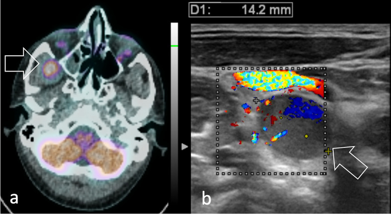
Figure 13.
26-year-old male diagnosed with pleomorphic head and neck RMS. (a) 11C-Methionine PET/CT shows the primary tumour as a round area of intense radiopharmaceutical uptake in the temporal fossa (arrow). (b) Color flow Doppler ultrasound of the neck shows a right lateral cervical lymph node with abnormal morphology, echogenicity and vascularization consistent with regional nodal involvement (arrow). RMS, rhabdomyosarcomas.

