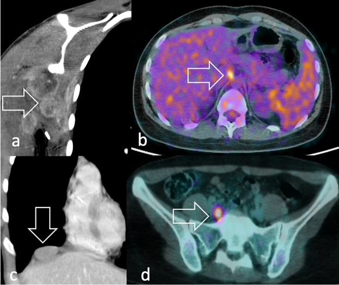
Figure 15.
Coronal post-contrast CT scan detects (a) multiple, confluent axillary lymphadenopathies and (c) right diaphragmatic adenopathy (arrow) in a patient with aRMS. 18F-FDG PET identifies (b) a tiny precrural and (d) retroperitoneal lymph node metastasis (arrow) in a 17-year-old patient diagnosed with nasal eRMS. 18F-FDG, 18F-fludeoxyglucose; PET, positron emision tomography; RMS, rhabdomyosarcomas.

