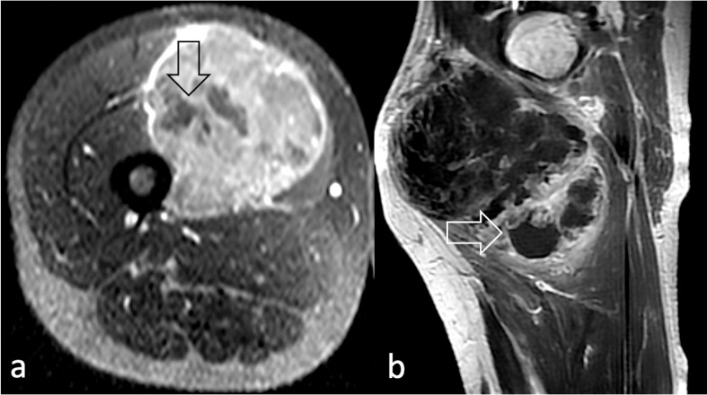
Figure 3.
Non-enhancing areas representing necrotic tissue (arrow) on post-contrast T1 W image in (a) post-contrast axial T1W image acquired at the level of the left thigh in a 39 year-old male diagnosed with pleomorphic RMS; (b) sagittal post-contrast T1 weighted image of the right thigh of a 49-year-old male diagnosed with spindle cell/sclerosing RMS. RMS, rhabdomyosarcomas; T1W, T1 weighted.

