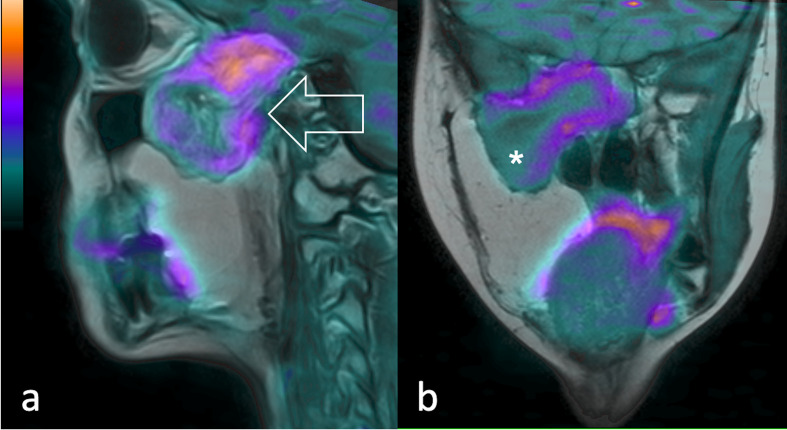
Figure 4.
19-year-old patient diagnosed with recurrent pleomorphic RMS. 11C-Methionine PET fused with T2 weighted MR images shows (a) in the sagittal and (b) coronal view of the lesion extending through the inferior temporal gyrus, middle cranial fossa, sellar region, masticatory space and pterygopalatine fossa (arrow). A central necrotic area is visible (asterisk). PET, positron emision tomography.

