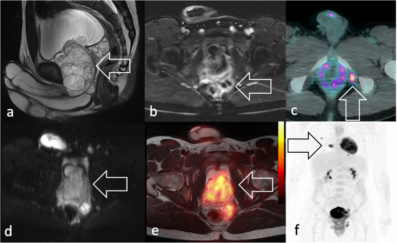
Figure 5.
Same 19-year-old patient of Figure 2a–b, diagnosed with eRMS. MRI shows in (a) sagittal T2 weighted image a voluminous, heterogeneous mass in the prostatic lodge (arrow) with (b) increased peripheral vascularisation seen in axial fat suppressed post-contrast T1 weighted image. The patient presented with (c) 18F-FDG PET/CT showed an isolated radiopharmaceutical uptake consistent with lymphadenopathy (arrow) and (d) marked signal restriction on axial DWI scan (e) 18F-FDG PET/T2W-MRI shows intense uptake at the level of the mass and multiple lung metastases in maximum intensity projection coronal plane (arrow). 18F-FDG, 18F-fludeoxyglucose; DWI, diffusion-weighted imaging; PET, positron emision tomography; RMS, rhabdomyosarcomas; T2W, T2 weighted.

