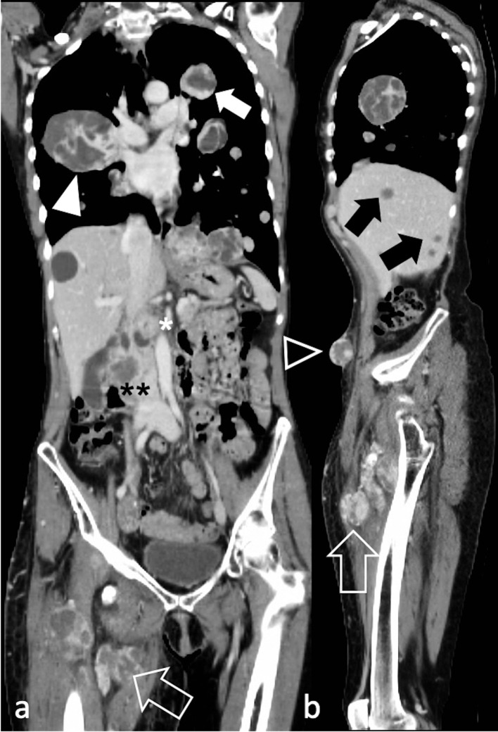
Figure 6.
Diffuse metastatic spread in a 60-year-old female with spindle cell/sclerosing RMS of the right thigh (arrow). Post-contrast CT depicts in a) coronal and b) sagittal reconstructions lung metastases (arrow), right hilar adenopathy (arrowhead), hepatic metastases (black arrows), pancreatic metastases (white asterisk), right renal metastases (black asterisks), and tumour deposits in the anterior abdominal wall (black arrowhead). RMS, rhabdomyosarcomas.

