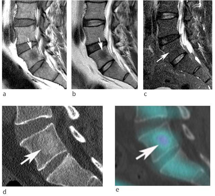
Figure 1.
53-year-old male being investigated for low back pain. (a) Sagittal T1W TSE, (b) T2W FSE and (c) STIR MR images demonstrate a poorly defined spherical lesion with reduced T1W and T2W SI (arrows) in the centre of the L5 vertebral body. The lesion is not identified on STIR. Note the absence of peri-lesional oedema (arrow-c). (d) Sagittal CT MPR demonstrates mild medullary sclerosis at the site of the lesion (arrow). (e) Sagittal SPECT-CT MPR demonstrates mild increased activity (arrow).

