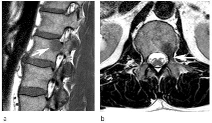
Figure 12.
A 50-year-old male referred for investigation of a lesion in the L1 vertebra. (a) Sagittal T1W TSE and (b) axial T2W FSE MRIs demonstrate a lesion in the left posterior third of the vertebral body extending into the pedicle (arrows). The SI characteristics are classical for FNMH. The lesion remained stable over 6 months.

