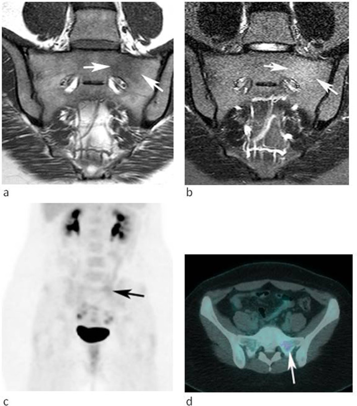
Figure 5.
A 28-year-old female referred for further investigation of a marrow abnormality in the left sacral ala found incidentally. (a) Coronal oblique T1W TSE and (b) STIR MR images demonstrate a poorly defined lesion which is slightly hyperintense to muscle on T1 and mildly hyperintense to marrow on STIR (arrows). (c) Coronal FDG-PET MIP and (d) axial-fused PET-CT images demonstrate mild increased activity. The lesion was stable over 25 months.

