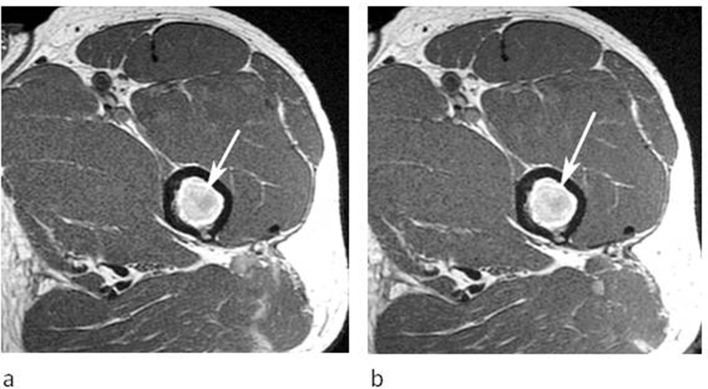
Figure 8.
A 43-year-old male referred for further investigation of a marrow abnormality identified in the left proximal femur. (a) Axial T1W TSE and (b) post-contrast T1W TSE MR images demonstrate a round lesion which is hyperintense to muscle on T1 (arrow-a) and shows no enhancement following contrast (arrow-b).

