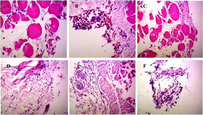
Fig. 1.
Histopathological analysis of rat synovium tissue. a Control group and b model group were treated with saline for 5 days. c High dose group d middle dose group and e low dose group were treated with extract of RDN for 5 days, dose amounted to 40 mg/kg, 80 mg/kg and 160 mg/kg daily, respectively. f COL group was treated with COL for 5 days. Each slide was examined at a magnification of 200 times. Histopathological changes of H&E-stained sections were used to assess the severity of GA, characterized by infiltration of inflammatory cells and synovium swelling and distention as well as vasocongestion and tissue necrosis. In contrast, healthy synovium showed almost no inflammatory cells

