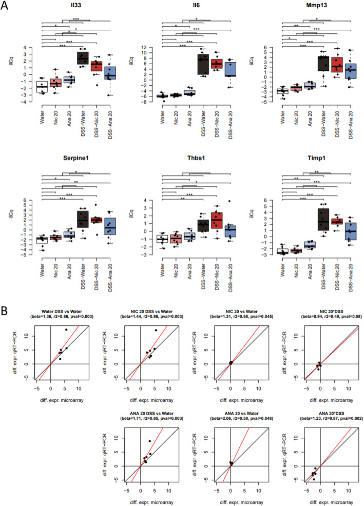
Fig. 4.
Validation of microarray transcriptomic results using qPCR. a Box-and-whisker plots for the distribution of the qPCR expression levels (δCq) of the selected genes. The boxes represent the quartiles while the whiskers extend to the most extreme data point which is no more than 1.5 times the interquartile range from the box. The horizontal brackets indicate the statistical significance of the corresponding comparisons (*,**,*** mean p-value smaller than 0.05, 0.01, 0.001, respectively). b Scatter plots comparing the mouse colon differential expression values obtained by microarray (horizontal axis) and qPCR (vertical axis). The following similarity metrics were indicated: “beta” is the slope of the best intercept-free linear fit between microarray and qPCR values, “r2” is the coefficient of determination measuring the “goodness-of-fit”, and “pval” is the p-value associated to the null hypothesis beta = 0. Nic 20, nicotine 20 mg/kg; Ana 20, anatabine 20 mg/kg

