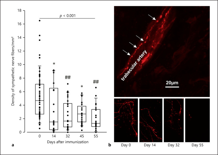
Fig. 1.
Density of sympathetic nerve fibers in the spleen of control and arthritic rats. a Density of sympathetic tyrosine hydroxylase-positive nerve fibers. Each dot represents the result for 1 mouse (mean from 10–17 high-power fields). * p < 0.05, ## p < 0.005 versus control 8 (day 0). The pvalue above the boxes gives the result from the ANOVA on ranks test for all groups. Data are given as box-plots with the 10th (whisker), 25th, 50th (median), 75th, and 90th (whisker) percentiles. b Immunohistochemical staining of sympathetic nerve fibers. In the top panel, a typical bead chain staining of a splenic artery is given. The arrows point to varicosities along the nerve fiber.

