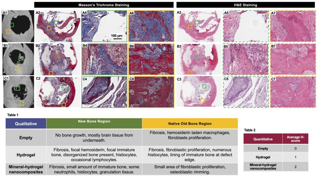
Figure 4: Histological evaluation of mouse critical sized defect area 12-week after implantation of mineral-hydrogel nanocomposites.
(A1, B1, and C1) μCT images of empty defect control, lyophilized CHT-KCA hydrogels, and lyophilized CHT-KCA mineral-hydrogel nanocomposites implanted mice used for histology sectioning, respectively. (A2, B2, and C2) Masson’s Trichro e stained stitched images of longitudinal section of empty defect control, lyophilized CHT-KCA hydrogels, and lyophilized CHT-KCA mineral-hydrogel nanocomposites implanted mice, respectively; (A3, B3, C3) Similar H&E stained stitched image of longitudinal section of empty defect control, lyophilized CHT-KCA hydrogels, and lyophilized CHT-KCA mineral-hydrogel nanocomposites, respectively; (A4-A5, B4-B5, C4-C5) Magnified images from new bone region (NB) are indicated in green boxes for control, hydrogels, and mineral-hydrogel nanocomposites, respectively. (A6-B7, C6-C7, D6-D7) Similar images from the native old bone region (OB) are shown in yellow boxes. Solid lined boxes are used to indicate Masson’s Trichro e stained i ages and dotted lined boxes are used for H&E stained images. (Table 1) Qualitative evaluation of H&E and Masson’s Trichro e stained longitudinal sections of mice calvaria from two different regions (Green-NB and Yellow-OB). (Table 2) Healing score (H-score) of the same sections from NB area; Score range: 0–4; NB- new bone, OB- old native bone.

