FIG 1.
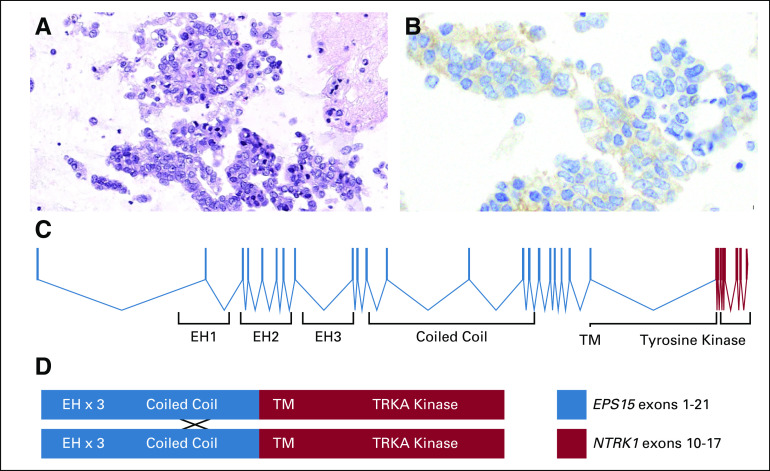
Pathologic and molecular features of a TRK fusion-positive lung cancer. Photomicrographs of (A) cytology from the patient’s lung adenocarcinoma and (B) positive immunohistochemistry results remarkable for cytoplasmic and membranous staining using a pan-TRK antibody are shown. (C) A map of the EPS15-NTRK1 fusion protein that resulted from an in-frame fusion between EPS15 exon 21 and NTRK1 exon 10. (D) A schematic that shows the domains of the fusion protein depicted as possibly dimerizing at the coiled-coil region of EPS15 where that protein is known to form homodimers.

