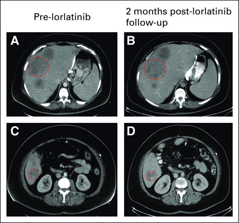
FIG 3.
Computed tomography imaging of hepatic metastasis before and after lorlatinib. In row 1, the circled hepatic lesion after disease progression on fluorouracil, folic acid, oxaliplatin, and irinotecan (A) before lorlatinib was stable in size (B) 2 months after lorlatinib treatment. Similarly, row 2 shows a different metastatic hepatic lesion (C) before lorlatinib with stable disease (D) after 2 months of lorlatinib. The patient currently continues to be treated with lorlatinib.

