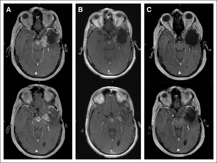
FIG 1.
Magnetic resonance imaging (MRI), axial postgadolinium T1-weighted imaging. (A) Baseline brain MRI images, before starting dabrafenib and trametinib (D+T), demonstrated progression of disease into the surgical cavity, as well as new lesions in the left insular region and midbrain. (B) Brain MRI images at 1 month after initiation of D+T, confirming > 50% decrease of all measurable enhancing lesions. (C) Brain MRI images at 7 months of D+T, demonstrating disappearance of all enhancing disease.

