
Figure 4.

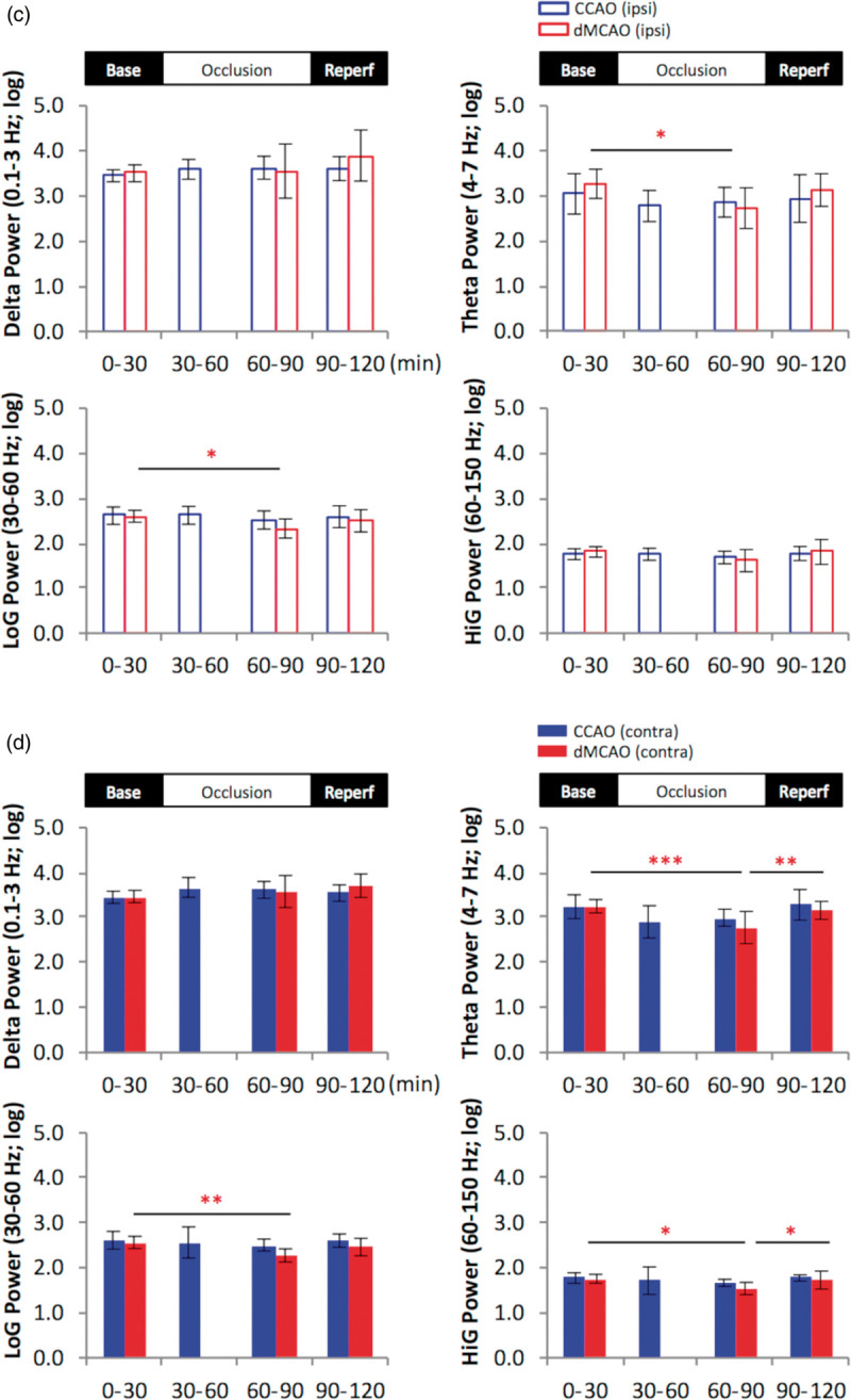
Cortical stroke induces temporary reduction in the power of theta and gamma oscillation in the hippocampal slm. (a and b), representative 2-h time series for the parameters as indicated in rats subjected to temporary cortical stroke (dMCAO) or mild ischemia (CCAO) for ipsilateral (a) and contralateral (b) recordings in the slm. Data are expressed in logarithm scale. Black lines and gray shades represent mean and SD, respectively. (c and d), Quantification of temporal changes in hippocampal oscillatory activity in ipsilateral (upper open bar panels) (c) and contralateral hippocampus (lower solid bar panels) (d). The continuous data for each variable were grouped into four time periods as baseline (0–30 min), 1st occlusion (30–60 min), 2nd occlusion (60–90 min) and reperfusion (90–120 min). One-way analysis of variance (ANOVA) for the time periods was used to determine changes induced by CCAO (blue) or dMCAO. Data for the 1st occlusion period in the dMCAO group were only partially plotted or excluded for statistics due to a procedural delay. A follow-up post hoc analysis was performed to reveal a detailed temporal profile for each variable if necessary. Theta and low gamma (LoG) power was reduced in the bilateral stratum lacunosum moleculare during dMCAO occlusion compared to baseline, while high gamma (HiG) power was reduced contralaterally. Unlike dMCAO, mild ischemia via CCAO did not induce significant changes in theta or gamma oscillations compared to baseline. The time series plots show greater dynamic changes of power in all frequencies during dMCAO reperfusion not revealed by the averages of power during the time period *p < 0.05, **p < 0.01, ***p < 0.001. dMCAO: N = 10; CCAO: N = 7.