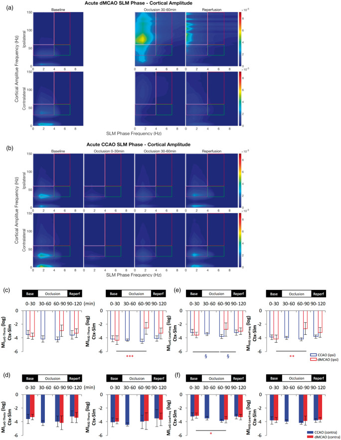
Figure 7.
Cortical gamma power is modulated by hippocampal low frequency phase in the slm following cortical stroke. Comparison of temporal modulation of cortical gamma amplitude by low frequencies oscillations phase in the slm layer of the ipsilateral and contralateral hemisphere revealed an increase of the theta-high gamma modulation index (a–d) and an increase of the coupling between high gamma and lower frequencies (0–4 Hz) (e–f) in the ipsilateral hemisphere during acute dMCAO. The phase-amplitude comodulogram (a–b) confirmed the coupling between theta or low frequencies and high oscillations observed in the bar graph representations (c–f) in the ipsilateral hemisphere during MCA occlusion. In contrast, little change in theta-gamma modulation or lower frequencies coupling was observed following CCAO except for a brief decrease of MiHiG-Theta (c) and MiLoG-Low Freq (e) during the CCA occlusion in the slm layer of the ipsilateral side. *p < 0.05, **p < 0.01, (for dMCAO); §p < 0.05 (for CCAO) for multiple comparisons between baseline and other levels. dMCAO: N = 10; CCAO: N = 7.

