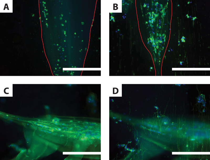
Figure 5.
Neurite Elongation in Composite Scaffolds. Immunofluorescent micrographs of primary cortical neurons 3 days post-encapsulation in IPC fiber / electrospun fiber composite scaffolds. Neurons are labeled with the neuron-specific marker tubulin-Tuj1 (green) and cell nuclei with DAPI (blue). A-B. Parallel composite scaffolds, where cell-encapsulating IPC fibers (outlined in red) were drawn such that the nuclear fiber bundle shared the same orientation as the underlying electrospun fibers. When IPC fibers were made using AsC (A), no neurites formed. When IPC fibers were made using WsC (B), encapsulated cells extended neurites unilaterally, as did de-encapsulated cells in contact with electrospun fibers. C-D. Two different focal planes of the same perpendicular composite scaffold containing IPC fibers formed using WsC (n = 3). Cells encapsulated in IPC fibers extend neurites horizontally, along the length of the nuclear fiber bundle (C). Neurites also extend vertically, along the underlying electrospun fiber mat (D). Similar images were collected from preparations in at least one other separate trial. Scale bars: 200 μm.

