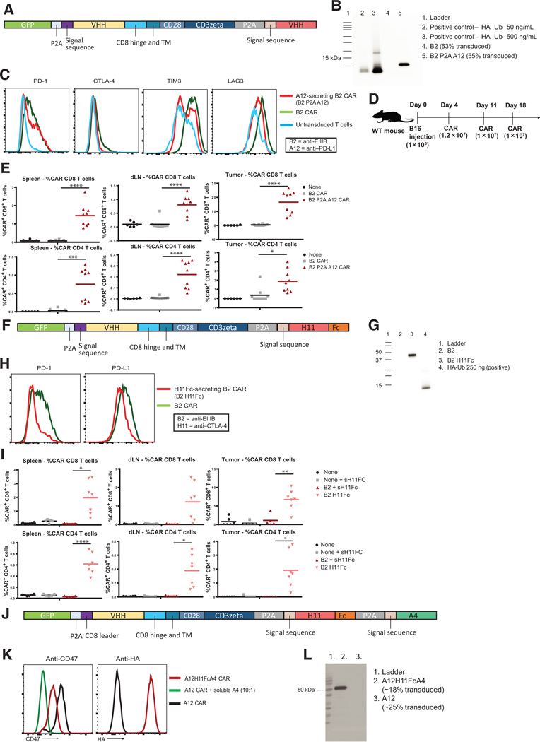
Figure 4.
Secretion of VHHs and VHH-fusions by CAR T cells is modular, and multi-cistronic constructs can be produced. A, Vector design of A12 VHH-secreting B2 CAR T cells. B, Anti-HA IP on supernatant from the culture of B2 CAR T cells or B2 A12-secreting CAR T cells. TM, transmembrane. C, Flow cytometry for common exhaustion markers in populations of untransduced cells and cells transduced with B2 CARs or B2 A12-secreting CAR T cells. D, Experimental schematic to determine how A12 secretion affects persistence of B2 CAR T cells. E, Flow cytometry to determine the presence of CD8+ and CD4+ CAR T cells in the spleen, tumor-draining lymph nodes, and tumors (spleen CD4, P = 0.0001; spleen CD8, P < 0.0001; draining lymph node CD4, P < 0.0001; draining lymph node CD8, P < 0.0001; tumor CD4, P = 0.0042; tumor CD8, P < 0.0001). dLN, draining lymph node. F, Vector design of anti–CTLA-4 H11Fc-secreting B2 CAR T cells. Experiments were repeated twice. TM, transmembrane. G, Anti-HA IP on supernatant from cells transduced with either B2 CAR or B2 H11Fc-secreting CAR. Samples are probed with an anti-HA-HRP–labeled antibody. H, Staining for common exhaustion markers on populations of cells transduced with either B2 CAR or B2 H11Fc-secreting CARs. I, Following a similar experimental setup as in D, we analyzed the persistence of B2 and H11Fc-secreting B2 CAR T cells in the spleen, draining lymph nodes (dLN), and tumors. The B2 H11Fc CAR T cells showed improved survival over B2 CAR T cells or B2 CARs given systemic doses of H11Fc (spleen CD4, P < 0.0001; spleen CD8, P = 0.0058; dLN CD4, P = 0.0053; dLN CD8, P = 0.0254; tumor CD4, P = 0.0088; tumor CD8, P = 0.0019). Experiments were repeated twice. J, Vector design of a multi-cistronic construct to generate CAR T cells with a VHH-Fc fusion and an additional VHH on a single plasmid. TM, transmembrane. K, Staining with anti-CD47 shows inhibition of binding. Staining with an anti-HA mAb shows production of HA-tagged VHH bound to the T-cell surface, indicating that A4 VHH is being produced. L, Anti-HA IP on supernatant from cells transduced with an H11Fc- and A4-secreting CAR T cell shows that H11Fc is secreted. *, P ≤ 0.05; **, P ≤ 0.01; ***, P ≤ 0.001; ****, P ≤ 0.0001.

