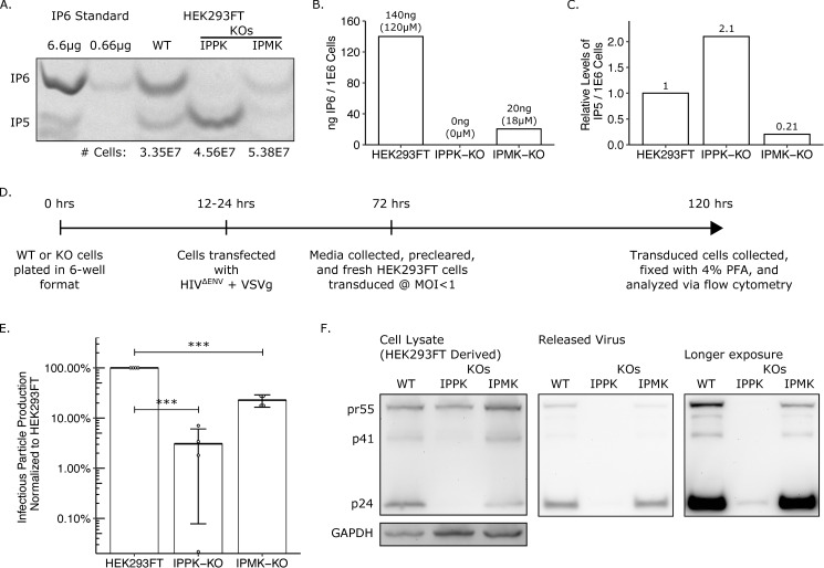
Fig 2. IP pathway KOs have reduced IP6 and IP5 levels and have a loss of infectious particle release.
(A) 33% PAGE gel separating inositol phosphates. Two dilutions of purified 1 M IP6 were used as a standard and had IP5 breakdown products. The number of cells in each sample is indicated. (B) IP6 quantification of panel A in ng per million cells and μM. (C) Relative IP5 quantification normalized to the HEK293FT control. (D) Experimental timeline. (E) Percent infectious particle release normalized to HEK293FT cells. Student’s t-test was used for pair-wise comparison (n = 4, *** p < 0.001, error bars = mean + SD). (F) Representative western blot of experiments from panel D. Full-length HIV-Gag (pr55) and GAPDH loading control are presented on the left panels. Virus released into media is presented in the middle panel. A longer exposure of the virus release blot was also taken and presented on the right panel.

