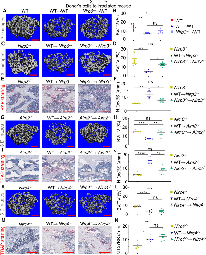
Fig 1. Radiation damages bones through the NLRP3 and AIM2 inflammasomes but not the NLRC4 inflammasome.
Three-month-old WT, Nlrp3-/-, Aim2-/-, and Nlrc4-/- male mice were left untreated or subjected to 9-Gy TBI. Irradiated mice were transplanted with 107 bone marrow cells from 3-month-old WT or null male mice to generate WT→WT, Nlrp3-/-→WT, WT→Nlrp3-/-, Nlrp3-/-→Nlrp3-/-, WT→Aim2-/-, Aim2-/-→Aim2-/-, WT→Nlrc4-/-, and Nlrc4-/-→Nlrc4-/- mice. The femurs were harvested 3 weeks later and analyzed by μCT. (A, C, G, K) Cross sections of 3D reconstructions. (B, D, H, L) BV/TV. The femurs were also stained for TRAP activity. (E, I, M) TRAP+ cells (OCs), stained in red. (F, J, N) N.Oc/BS. The numerical values underlying Fig 1B, 1D, 1F, 1H, 1J, 1L, 1N can be found in S1 Data. Data are mean ± SEM. *P < 0.05, **P < 0.005, ***P < 0.0005, ****P < 0.0001. Scale bar: 200 μm. μCT, micro–computed tomography; BV/TV, bone volume/total volume; N.Oc/BS, OC number/bone surface; ns, not significant; OC, osteoclast; TBI, total body irradiation; WT, wild-type.

