
Figure 3.
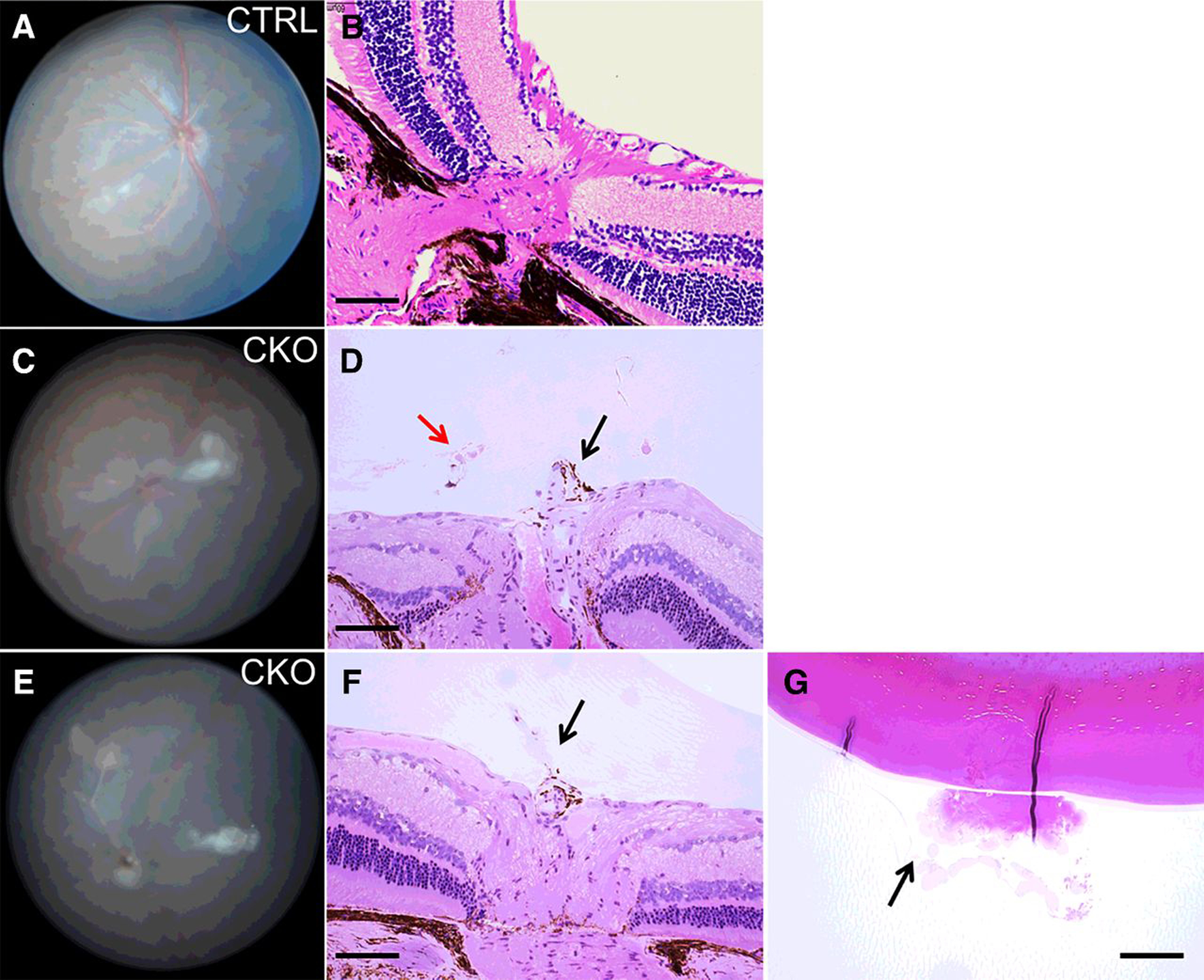
Histologic analysis of Vhl-mutant mice reveals PFV. A and B, Funduscopic and histologic images of a 5-month-old control mouse. The hyaloid artery has completely regressed. C and D, Representative funduscopy and histologic images of a 5-month-old Vhl-mutant mouse. Black arrow, persistent hyaloid vessel; red arrow, retinal neovascularization in the vitreous. E–G, Another Vhl-mutant mouse of similar age exhibited persistent hyaloid vessel (F, arrow), and vitreous fibro-neovascular structure adherent to the lens (G, arrow). Scale bars for B, D, F, G, 100 μm.