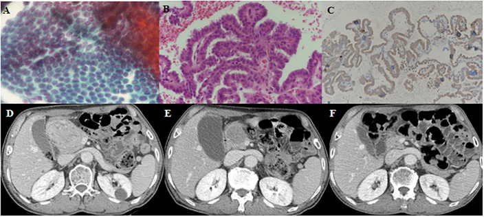
Fig 2. A 71-year-old male patient presenting with pancreatic metastasis from papillary thyroid cancer.
(A, B, C) Endoscopic ultrasonography-guided fine-needle aspiration (FNA) and biopsy showed metastatic PTC on the pancreatic head mass. (A): Hematoxylin and eosin (H-E) staining of FNA (x 400), (B): H-E staining of biopsy (x400), (C): BRAFV600E staining, (D) Abdomen computed tomography (CT) showed 6.9 cm heterogeneously enhancing mass on the pancreatic head before lenvatinib therapy. (E) Follow-up abdomen CT at 3 months after lenvatinib therapy showed the pancreatic tumor decreased in size from 6.9 cm to 4.1 cm. (F) Follow-up abdomen CT at 24 months after lenvatinib therapy showed that the pancreatic tumor was significantly reduced to 1.2 cm.

